Microfluidics and Bioimaging Lab
Microfluidics and Bioimaging Laboratory at UofSC: facilities
Microfluidics and Bioimaging Laboratory is equipped with cutting-edge and advanced instrumentations for fundamental and applied research in biomedical engineering, micro/nanofluidics, low Reynolds number (bio)fluid dynamics, Electrokinetics and interfacial flows. This well-equipped state-of-the-art laboratory has over 1000 square feet.
The Micro/Nanofluidics Lab nanoscopic system. Partial view of the femtosecond laser based STED nanoscopy and multimodality bioimaging system in the University of South Carolina.
Optical Instruments
-
Laser scanning nano/microscope 3-color (in-house developed)
-
Femto lasers based far field Nanoscopy: multifunctional far-field nanoscopic system, which is consisted of Coherent’s Chameleon Ultra II 80MHz (RoHS) Ti:Sapphire tunable femto laser and Mira OPO – Fan Poled Ring Configuration optical parametric oscillator (OPO), Nikon’s C2 laser Scanning Confocal Microscope System, and PI’s PInano XYZ P-545.3C7 Piezo Stage with Capacitive Sensors with USB controller for velocity, concentration and temperature measurement, nanofabrication and bioimaging of live cell. The system has the following multimodality imaging functions:
-
Stimulated Emission Depletion (STED)
-
Multiphoton (PM)
-
Fluorescence life time imaging (FLIM) (Becker & Hickl GmbH)
-
Fluorescence resonant energy transfer (FRET)
-
Second harmonic generation microscope (SHGM)
-
Femtosecond laser-based laser induced fluorescence photobleaching anemometer (LIFPA)
-
CW laser STED system for far field nanoscopic bioimaging system and LIFPA
-
405nm laser (Crystalaser)
-
2Watt 532nm diode pumped solid state laser (OEM laser system)
-
-
-
Nikon 3 color C2 laser scanning confocal microscope
-
Diode laser 405 nm 20 mW
-
300 mW 473 nm laser
-
He-Ne lasers 633 nm (17 mW, Newport Inc.)
-
-
Epifluorescence and other microscope
-
Olympus inverted epifluorescence microscopes (IX70, Olympus)
-
Olympus: PLan APO 1.5X; PL APO 4X; 10X; PL APO 20X; PL APO 40X; PL APO 100X /NA 1.4 oil.
-
-
-
LaVision Particle Imaging Velocimetry (PIV)
-
LaVision Particle Imaging Velocimetry system with Nd:YAG Dual Cavity pulsed laser - New Wave Research Solo PIV III 15, 2 x 50 mJ/pulse @ 532 nm, 15 Hz pulse rate
-
-
Microscope objective
-
Leica: HCX PL APO 100X/1.4; PL APO 63X/1.32; PL APO 40X/1.25; HCX PL APO 40X/0.85; HC PL APO 20X/0.7; HC PL APO 10X0.4 and HC PL Fluotar 5x/0.15 ∞/-/D
-
Nikon: PL APO 4X; PL APO 20X; PL APO 63X/1.4; PL APO 100X/1.4 VC
-
-
Detector
-
Autocorrelator FR-103MN (Femtochrome Research Inc)
-
Ultra-low noise single photon detection module (id100-MMF50, Becker & Hickl Inc. Germany)
-
Horiba Jobin Yvon H-10 Monochromator H-10vis
-
Multiple photomultipliers tube (R6060; R1464,) from Hamamatsu
-
The Micro/Nanofluidics Lab clean room.
-
Camera:
-
Tow Cooke SensiCam QE Cameras (high speed, Cooke corp.), one Qimage Fast 1394 and a HDCE-30A.
-
-
Opto-mechanical parts
-
Physik Instrument’s PInano XYZ P-545.3C7 Piezo Stage with Capacitive Sensors and USB controller
-
Physik Instrument’s Piezo Nano 3D positioning Stage (E664, PI)
-
Two Picomotor Piezo Mirror Mounts
-
Multiple 3D Translation stages (Newport, Thorlabs, Mellesgriot)
-
Multiple lenses and optical filters, mechanical parts and etc.
-
-
Anti-vibration optical tables (Thorlabs)
Electronic test instruments
-
Oscilloscope (4 GS/s, 4 channels, LeCroy 9384CL), Oscilloscope (BK Precision)
-
Lack-in amplifier from Stanford Research
-
The HVS448 High Voltage Sequencer, LabSmith
-
Digital delay / pulse generator (DG535, Stanford Research System)
-
Keithley 248 high voltage power supply (Keithley Instruments Inc)
-
Two Micro driver controller (Newport)
-
High voltage power supply (0~8000v, 5 channels) (EMCO Inc.)
-
DC power supply (6634B, Agilent)
-
Multimeter (34401A, HP )
-
AD Convertor (USB-6259, NI)
-
Two AFG3102 function generators from Tektronix
-
Low-noise current preamplifier SR570 (Stanford Research System)
General lab equipments
-
3 Cell Culture CO2 Incubators (Sanyo)
-
Fume hood
-
2 PHD Syringe pumps (Harvard apparatus)
-
Branson ultrasonic cleaner (BRANSONICR )
-
vortex mixer (Fisher Scientific),
-
Electronic weighing balances (METTLER AT261 DeltaRangeR ,Resolution: 0.01mg)
-
Vacuum pump Pascal type 2010 (Alcatel vaccum products Inc.)
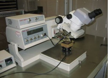
The Micro/Nanofluidics Lab Wire Bonder .
-
PC-410 Magnetic Stirrer (CorningR)
Microfabrication system
-
Clean room, class 1000
-
Wire Bonder (Westband)
-
Headway Research CB-15 Photoresist spinner
-
Plasma cleaner
-
Electric Oven (Apothecaries Sundries Mfg. Co.)
-
UV light source
Water channel for fluid dynamics and turbulence
Water channel
-
The water channel can be used for
-
Pipe flow
-
Mixing layer
-
Wake
-
Step flow
-
under active forcing
-
The system can be used for fluid mixing research
-
The PIV system and laser induced fluorescence are used for the water channel


I'm a paragraph. Click here to add your own text and edit me. It's easy.
Clean room of the Microfluidics & Bioimaging Lab
Wire Bonder of the Microfluidics & Bioimaging Lab
Water Channel of the Microfluidics & Bioimaging Lab